
Magnification Making the invisible visible


Magnification Objectives:
- Describe the magnification possible with hand lenses and microscopes.
- Explain why light sources and stains are important in microscopy.
- Provide a historical example of how microscopes changed the way people viewed the world.
When you think of a biology lab, what is the main piece of equipment you expect to see?
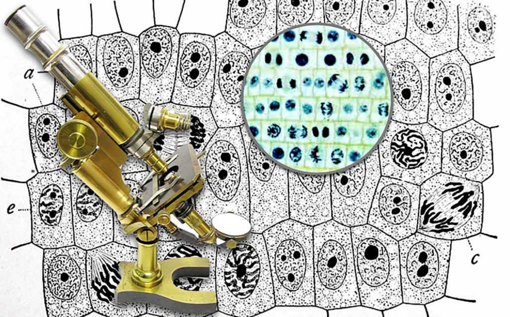
The earliest optical or “light” microscopes date back to the seventeenth century, magnifying specimens beyond the range of normal human vision.
Early magnification was approximately 10x, or ten times the unaided eye, now microscopes that shine light through specimens can magnify over 1500x and electron microscopes developed in the 20th century magnify 200,000x. Most of the research topics discussed in this course requiring microscopic examination, or “microscopy,” would use an optical “light” microscope.
A bright light shines through a thin tissue of cells and finely ground glasses magnify the image. Both the eye pieces and specialized lenses called “objectives” magnify. From this image, if you have a 10x eye piece and a 10x objective, what is the total magnification?
answer: 100x
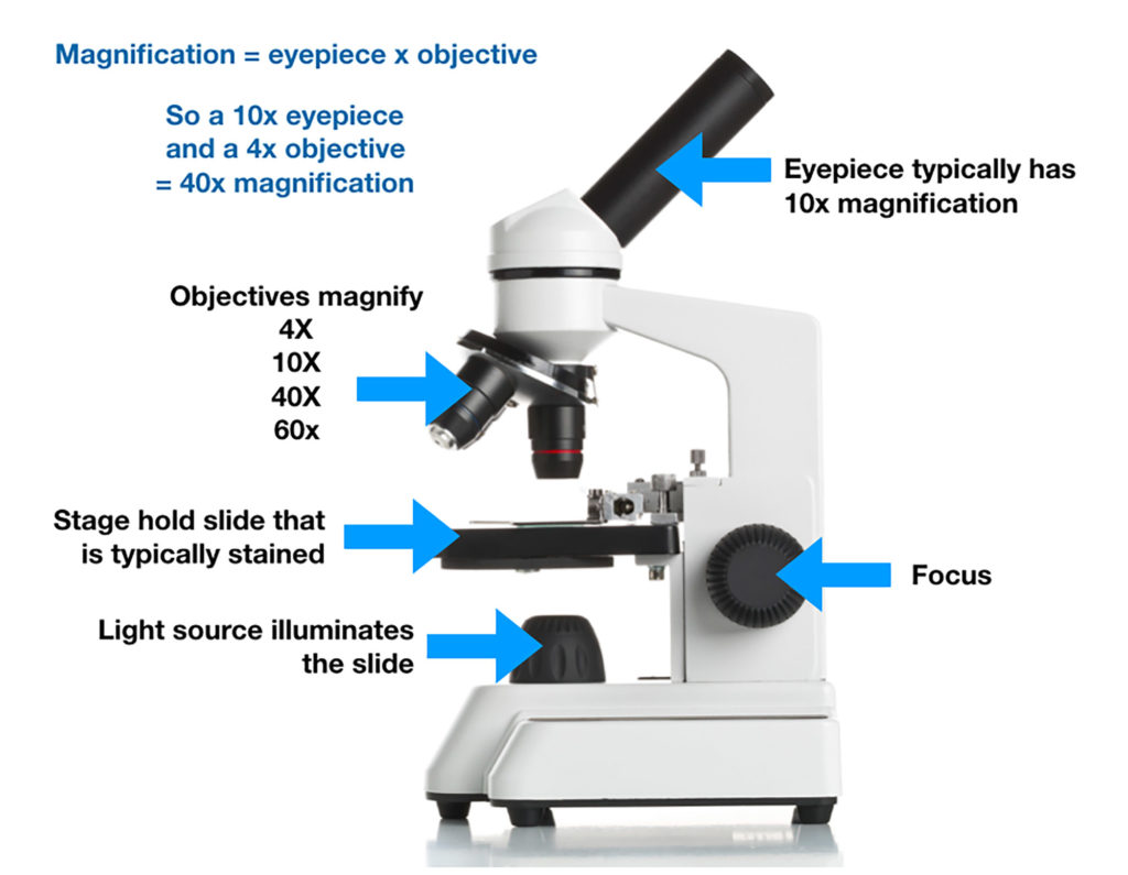
The bright light can make specimens look clear and colorless, so pigments or stains are often used to color certain parts of cells and the tissues they form.
Here are a few basics on glass lenses and light sources in microscopes, as well as staining of animal tissues in microscope slide preparation
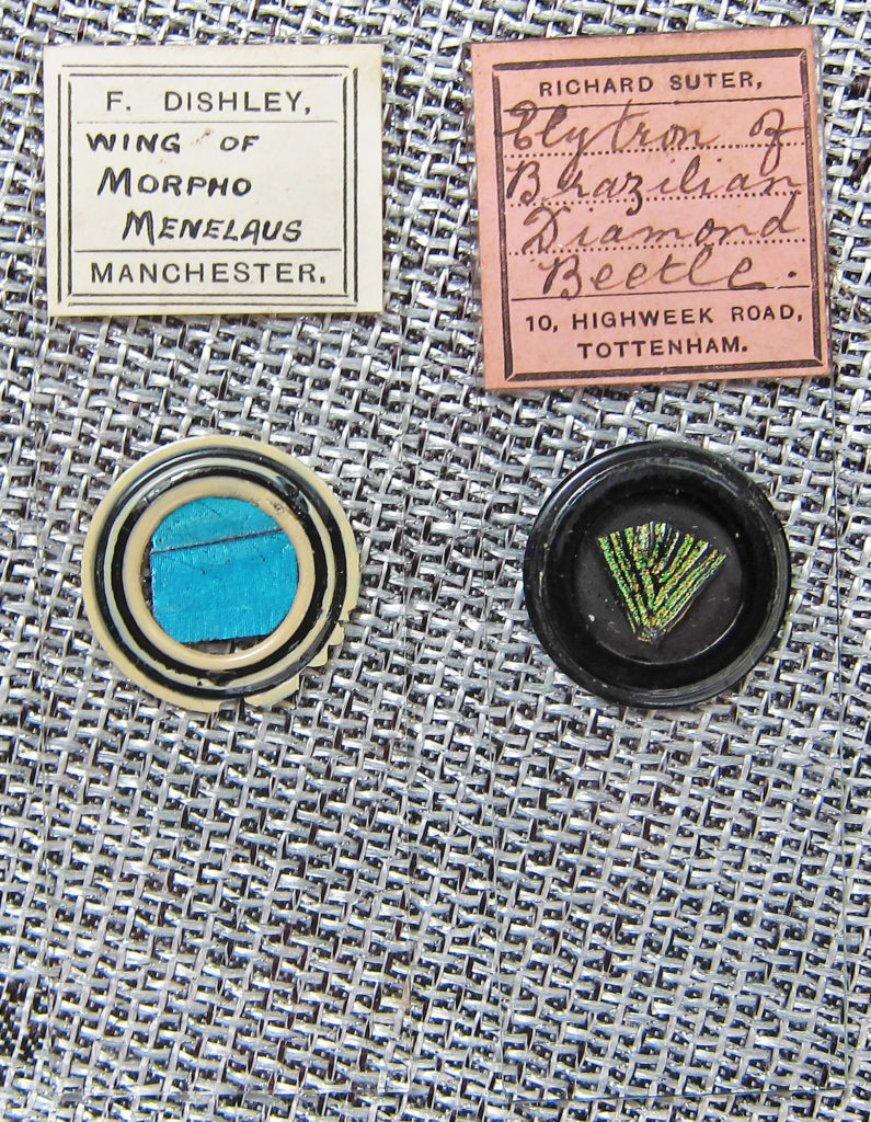
Antique microscope slides contain specimens of rare, sometimes extinct, organisms. They were also frequently prepared with chemicals that are now known carcinogens (cancer-causing agents), and are no longer used. Unfortunately the high quality coloring and contrast produced by these stains has not been fully replicated.
Stains can turn clear cells that are difficult to read into powerful sources of information. Using a medical example, women get regular pap smears to detect possible abnormal growth in the cells lining the cervix. These cells are colorless under bright light until stain is added.
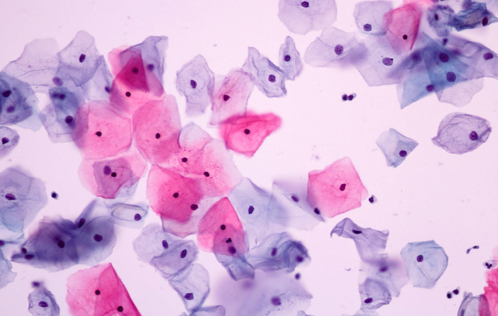
Normal Cells 200x
The normally colorless cells are staining shades of pink and purple, which indicates different stages of the cells. The pink were on the surface being sloughed off, the purple were a bit deeper in the lining tissue. The are all about the same size with similar size and shape nuclei (dark purple dots).
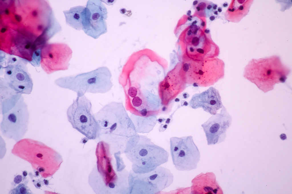
Abnormal Cells 200x
At first this may look similar, but look at the different colors and sizes of the nuclei in the cells. They are especially different in the bluer cells that are deeper in the cervical lining, This indicates abnormal growth and could not be easily detected without stains.
Microscopy has changed the way we experience the world
Following Darwin’s success aboard the H.M.S. Beagle, a science expedition was planned aboard the H.M.S. Challenger in 1875. The mission in part was to discover whether life existed in the depths of the oceans. Without light, it was thought that life could not exist.
The ship had a dredging mechanism to pull samples out of deep submersed ocean soils.
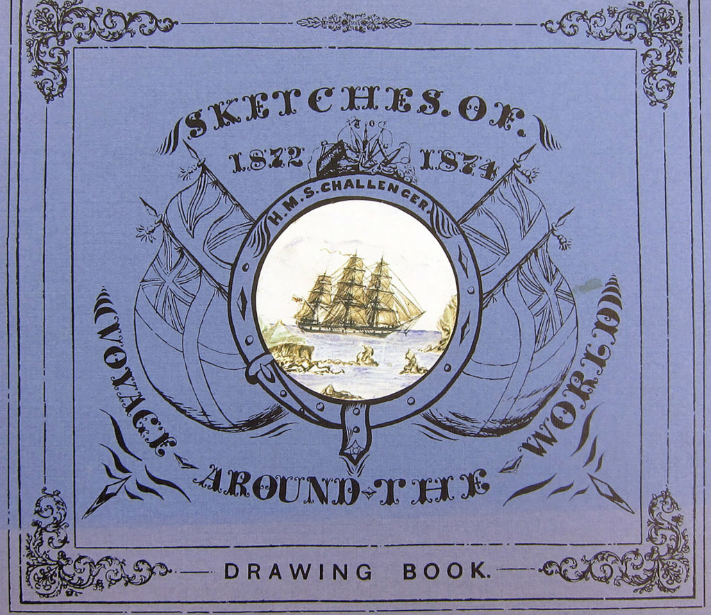
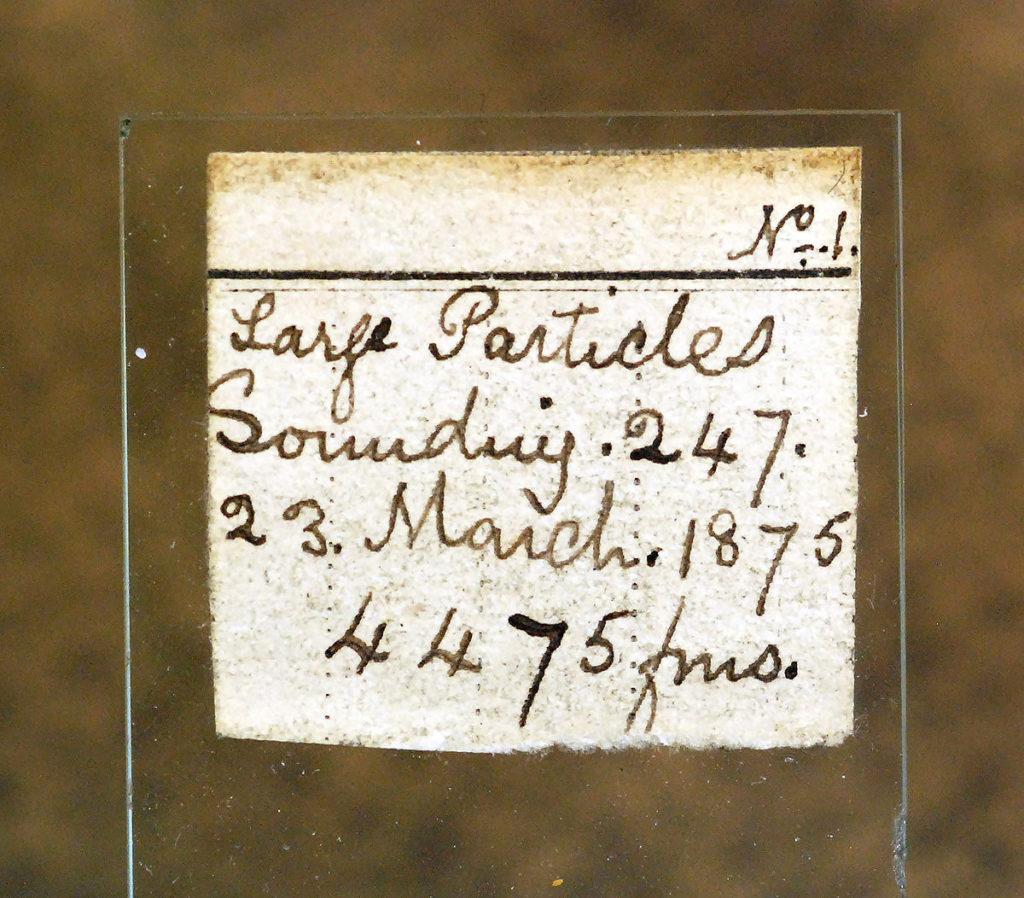
We have been fortunate enough to obtain two microscope slides prepared during this research voyage.
This sample was taken at 4475 fathoms, about 26,000 feet beneath the ocean surface, from an area of the Pacific Ocean now known as the “Challenger Deep,” named after this ship.
They are not stained, this is what the researchers saw on their own microscopes aboard the ship.
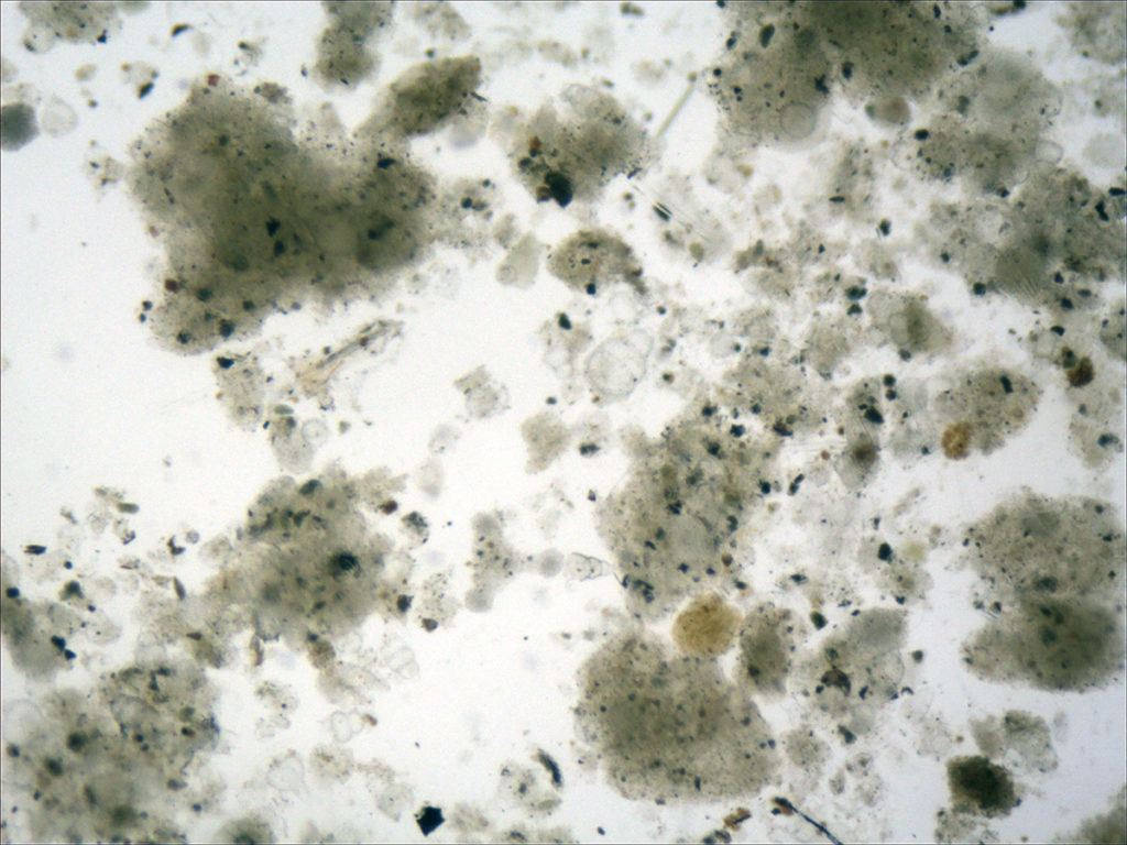
45x
At this lower magnification, the colors and clumps suggest the organization of organisms.
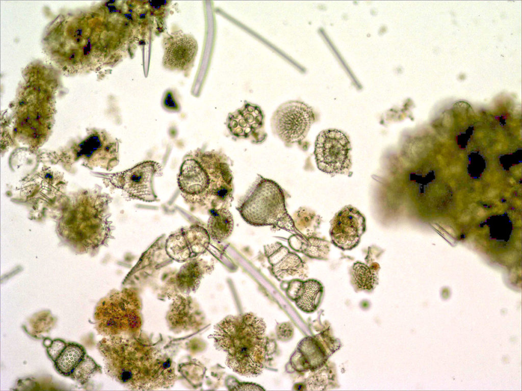
100x
There are definitely organisms, including algae (green), other protists, and small animals.
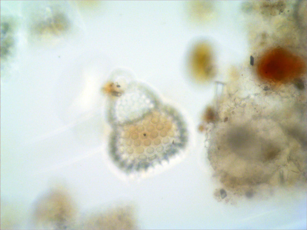
400x
A small protist, from the Kingdom that gave rise to animals and remains abundant in aquatic habitats.
Microscopy has played a critical role in the advancement of many fields, including medicine
You don’t need an expensive microscope to observe organisms. Here we are using a phone to magnify.
We will take a closer look at these pond samples in Module 7.
In the next section we will use microscopes to examine lichen communities.

Check your knowledge. Can you:
- Describe the magnification possible with hand lenses and microscopes?
- Explain why light sources and stains are important in microscopy?
- Provide a historical example of how microscopes changed the way people viewed the world?


